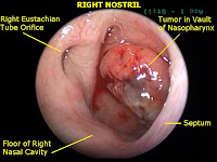
 Be carefull if you smoke ciggaret and eat saltfish too much, because they make deadly disease called Nasopharyngeal Carcinoma or Nasopharyngeal Cancer. Today Mbah Dukun Bagong, Indonesian Modern Shaman special author from Medical and Health Information explains about this cancer. wish it is so usefull for us.
DEFINITION
Carcinoma or cancer is a malignant growth of new cells, composed of epithelial cells that tend to infiltrate surrounding tissues and cause metastases.
Nasopharyngeal carcinoma is a malignant tumor arising in the epithelial coating the space behind the nose (nasopharynx).
ETIOLOGY
The link between Epstein-Barr virus and the consumption of salted fish said to be the main cause of this disease. Epstein-Barr virus can enter the body and remain there without causing an abnormality in a long time. To activate this virus requires a mediator. The habit of eating salted fish are constantly starting from childhood, is the main mediator that can activate the virus causing nasopharyngeal carcinoma.
Mediator below are considered influential to the onset of carcinoma
nasopharyngeal namely:
1. Salted fish, preserved foods and nitrosamines.
2. State of low socio-economic, environmental and lifestyle habits.
3. Frequent contact with substances that are considered carcinogens, such as:
- benzopyrenen
- benzoanthracene
- Chemical gases
- Industrial smoke
- Wood smoke
- Some plant extracts
4. Race and ancestry
5. Chronic inflammation of the nasopharynx
6. HLA profile
 |
| Saltfish causes nasopharyngeal carcinoma |
HISTOPATHOLOGIC
Microscopically, nasopharyngeal carcinoma can be divided into three forms, namely:
1. ulcerative forms
This form is most often found on the posterior wall and the area around the fossa rosenmulleri. Can also be found on the lateral wall in front of the tube and on the roof eustachius nasopharynx. These lesions are usually accompanied by smaller necrotic tissue and is very easy to conduct infiltration into surrounding tissue. Histopathologic picture is a form of squamous cell carcinoma with good differentiation.
2. nodular / lubuler / proliferative forms
Nodular form or lobuler very often found in the area around the estuary eustachius tube. This type of tumor shaped like grapes or polypoid rarely, found the existence of ulceration, but is sometimes found a small ulceration. Histopathologic picture of carcinoma usually without differentiation.
3. exophytic forms
exophytic forms usually grow on one side of the nasopharynx, found no presence of ulceration, stemmed and sometimes slippery surface. This type of tumor usually grows from the roof of the nasopharynx and can fill the entire cavity of the nasopharynx. These tumors can encourage the palate mole down and coana grow toward and into the nasal cavity. Histopathologic picture is a limphosarcoma
Classification of histopathologic picture of Nasopharyngeal cancer recommended by the Organization
World Health Organization (WHO) before the year 1991, divided into three types, namely:
1. Keratinizing Squamous Cell Carcinoma.
This type of differentiation can be subdivided into good, moderate and bad.
2. Non-keratinizing carcinoma.
In this type of differentiation is found, but no cell differentiation
Squamous intersel without bridges. In general, the cell boundary is quite clear.
3. Undifferentiated carcinoma.
In this type of individual tumor cells showed a vesicular nucleus,
oval or round with a clear nukleoli. Generally, the cell boundaries are not clearly visible.
Types without differentiation and without keratinization typically have the same are radiosensitive. While the type with keratinization is not so radiosensitive.
Histopathological picture of the latest classification recommended by WHO
in 1991, just divided into two types, namely:
1. Keratinizing Squamous Cell Carcinoma.
2. Non-keratinizing carcinoma.
This type can be subdivided into differentiated and not differentiated.
The main symptoms are:
1. Nasal sign:
· Colds that do not heal
· Epistaxis. Blood discharge is usually over and over again, few in number and often mixed with mucus, so it appears pink · Snot may like pus, watery or thick and smelly.
2. Ear sign:
· Tinnitus. Suppress tumor eustachii estuary causing tubal tubal occlusion, because the tube eustachii estuary, close to the fossa rosenmulleri. The pressure in the tympanic cavity is lowered, resulting in tinnitus.
· Conductive hearing loss
· Discomfort in the ear until the ear pain (otalgia).
3. Eye sign:
· Diplopia. Tumor crept laseratum foramen and cause disruption N. IV and N. VI. When exposed Chiasma optic would cause blindness.
4. Tumour sign:
Enlarged neck glands lymphoid · this is the spread or metastases near the lymphogen of nasopharyngeal carcinoma.
5. cranial sign
Cranial symptoms occur when the tumor has spread to the brain and is felt in people. These symptoms include:
· Constant headaches, pain is a metastasis in
haematogenous.
· Sensitibilitas of Regional cheeks and nose reduced.
· Difficulty on swallowing
· Aphonia
· Syndrome or syndrome reptroparotidean jugular Jackson on N. IX, N. X,
N. XI, N. XII. With signs of paralysis on:
o tongue
o palate
o Pharynx or larynx
o M. sternocleidomastoideus
o M. trapezeus
PATHOGENESIS and PATHOPHYSIOLOGY
1) Epstein-Barr Virus
Epstein-Barr virus replicate in epithelial cells and becomes latent in B lymphocytes Epstein-Barr virus infection occurs in two main places of salivary gland epithelial cells and lymphocytes. EBV infection of B lymphocytes initiate by binding to virus receptors, namely the complement component C3d (CD21 or CR2). Glycoprotein (gp350/220) to the capsule EBV binds to the CD21 protein on the surface of lymphocytes B3.
This activity is a series of chain starting from the entry of EBV into B lymphocytes and subsequent DNA causes B lymphocytes to be immortal. In the meantime, until now the mechanism of EBV entry into nasopharyngeal epithelial cells can not be explained with certainty. However, there are two receptors are thought to play a role in entry of EBV into epithelial cells of the nasopharynx and PIGR CR2 (Polimeric Immunogloblin Receptor). Cells infected by Epstein-Barr virus can cause several possibilities, namely: cell to die when infected with Epstein-Barr virus and virus replication conduct, or Epstein-Barr virus that can lead to cell death meninfeksi virus so that the cells return to normal or transformation can occur namely cell interactions between cells and viruses that result in changes in properties of the cell resulting in cell transformation into malignant cancer cells forming.
EBV genes expressed in patients with Nasopharyngeal Carcinoma is a latent gene, namely Ebers, EBNA1, LMP1, LMP2A and LMP2B. EBNA1 protein plays a role in maintaining the virus in latent infection. Transmembrane protein LMP2A and LMP2B inhibits tyrosine kinase signaling that is believed to inhibit viral lytic cycle. Among those genes, genes that were most responsible for cell transformation is the LMP1 gene. structure
LMP1 protein consists of 368 amino acids that is divided into 20 amino acids at the N terminus, six transmembrane protein segments (166 amino acids) and 200 amino acids at the carboxy end (C). LMP1 transmembrane protein mediates the signal for TNF (tumor necrosis factor) and improve the regulatory cytokine IL-10 which memproliferasi B cells and inhibit the local immune response.
2) Genetic
Although nasopharyngeal carcinoma tumor does not include genetic, but susceptibility to nasopharyngeal carcinoma in a particular community group has a relatively prominent and familial aggregation. Correlation analysis showed HLA (human leukocyte antigen) and cytochrome P450 enzyme gene pengode 2E1 (CYP2E1) is the possibility of gene susceptibility to nasopharyngeal carcinoma. Cytochrome P450 2E1 is responsible for metabolic activation of nitrosamines and related carcinogens.
3) Environmental factors
A large number of case studies conducted in populations residing in different regions in asia and north america, has confirmed that the fish sauce and other foods that contain large amounts of preserved nitrosodimethyamine (NDMA), Nitrospurrolidene (NPYR) and nitrospiperidine (NPIP), which may be a factor carcinogenic nasopharyngeal carcinoma. Also smoking and exposure to secondhand smoke who smoke cigarettes that contain formaldehyde and wood dust tepapar recognized risk factors for nasopharyngeal carcinoma by means of reactivating EBV infection.
DIFFERENTIAL DIAGNOSIS
1. adenoid hyperplasia
Usually found in children, rare in adults, in children
hyperplasia occurs Because repeated infections. On the plain will be seen a mass of soft tissue on the upper side of the nasopharynx generally demarcated and generally symmetrical and surrounding structures did not appear the signs look bleak infiltration in carcinoma.
2. Angiofibroma juenilis
Usually found in relatively young age with symptoms resembling nasopharyngeal carcinoma. The tumor is rich in blood vessels and the bias is not infiltrative. On the plain will get a mass on the roof nasofairng demarcated. The process can be extended seperrti on the spread of carcinoma, although rarely cause bone destruction
erosion simply because tumor suppression. Usually there is bending toward the front of the rear wall of the maxillary sinus is known as the antral sign. Because these tumors are rich in the vascular external carotid arteriography is necessary because the picture is very characteristic. Sometimes it is also hard to distinguish angiofibroma juvenils with nasal polyps on the plain.
3. Sinus tumors sphenooidalis
Primary malignant tumors sphenoidalis sinus is extremely rare and usually the tumor had reached somewhat advanced stage when the patient came to the first examination.
4. neurofibroma
Group of these tumors often arise in the lateral pharyngeal space that resembles a malignancy in the lateral wall of the nasopharynx. the C.T. Scan, pendesakan space medially toward the pharynx can help distinguish this group of tumors with nasopharyngeal carcinoma.
5. parotid gland tumor
Parotid gland tumors, especially those from the lobe that lies somewhat in the space of the pharynx and protruding towards the lumen of the nasopharynx. in most cases seen pendesakan parafaring space medial direction that appears on CT scan examination.
6. Chordoma
Although Chordoma is a major sign of bone destruction, but in view of nasopharyngeal carcinoma
too often cause bone destruction, it is often made it difficult to
membedakanya. With a plain, visible calcification or destruction, especially in the clivus region. CT can help to see if there is enlargement of the upper cervical glands because Chordoma typically do not pay attention to an abnormality in the gland, while nasopharyngeal carcinoma frequently metastasize to lymph nodes.
7. Menigioma cranial base
Although these tumors are somewhat rare, but the picture is sometimes resemble
nasopharyngeal carcinoma with sclerotic signs in the area of the cranial base. CT picture of meningioma is quite characteristic that is a bit hyperdense before injecting a contrast agent and will
become very hyperdense after administration of intravenous contrast agent. Arteriography examination also greatly aid in the diagnosis of this tumor.
STADIUM
Most recent staging based on an agreement between the UICC (Union
Internationale Contre Cancer) in 1992 are as follows:
T = Tumor, describes the state of the primary tumor, large and expansion.
T0: No visible tumor
T1: Tumor limited to one location in the nasopharynx
T2: Tumor extends more than one location, but still inside the cavity of the nasopharynx
T3: Tumor extends into the nasal cavity and / or oropharynx
T4: Tumor extends to the skull and / have about the brain's nerve
N = nodule, described the state of regional lymph nodes
N0: No enlargement of the gland
N1: There homolateral gland enlargement can still be driven
N2: There is enlargement of the gland contralateral / bilateral still be driven
N3: There is enlargement of both glands homolateral, contralateral or bilateral, which already
attached to the surrounding tissue.
M = metastases, distant metastases describe
M0: No distant metastasis
M1: There is distant metastasis.
Based on the above TNM, stage of disease can be determined:
Stage I: T1 N0 M0
Stage II: T2 N0 M0
Stage III: T3 N0 M0
T1, T2, T3 N1 M0
Stage IV: T4 N0, N1 M0
Any T N2, N3 M0
Any T Any N M12
According to the American Joint Cancer Committee in 1988, staging of tumors
nasopharynx are classified as follows:
Tis: Carcinoma in situ
T1: The tumor is found on one side of the nasopharynx or tumor that can not be seen, but
can only be known from the biopsy results.
T2: The tumor that attacks the two places, the wall of the postero-superior and lateral walls.
T3: tumor extension up into the nasal cavity or oropharynx.
T4: The tumor that spread to invade the skull or cranial nerves (or both).
PROGNOSIS
Overall, the 5-year survival rate was 45%. Prognosis is worsened by
several factors, such as:
· Stadium further.
Age of more than 40 years
· Men than women
· China than in whites
The enlargement of the gland neck ·
· Existence of damage to brain nerve palsy skull
· Presence of distant metastases
COMPLICATIONS
1. Petrosphenoid syndrome
The tumor grows upward into the base of the skull through the foramen laserum until sinus
cavernosal nerves pressing N. III, N. IV, N. VI also suppress N.II. which gives abnormalities:
· Trigeminal neuralgia (N. V): Trigeminal neuralgia is a pain in the
sesisi face marked with flavors such as exposed electrical flow is limited
on the distribution of the trigeminal nerve.
· Ptosis palpebra (N. III)
· Ophthalmoplegia (N. III, IV N., N. VI)
2. Retroparidean syndrome
The tumor grows forward toward the nasal cavity can then infiltrate into the surrounding. Tumor to the side and back toward the area where there retropharing parapharing and lymph nodes. The tumor is pressing against a nerve N. IX, N. X, N.
XI, N. XII with the manifestation of symptoms:
· N. IX: difficulty swallowing because of hemiparesis and the superior constrictor muscle
Taste disturbance at the rear third of the tongue
· N. X: hyper / hipoanestesi moles palate mucosa, pharynx and larynx with
respiratory disorders and salivary
° N XI: paralysis / atrophy bibs trapezius, SCM muscles as well as the palate hemiparese
mole
· N. XII: hemiparalisis and atrophy of the tongue side.
· Horner's syndrome: paralysis of the N. simpaticus servicalis, a narrowing of the fissure palpebralis, onoftalmus and miosis.
3. Cancer cells can contribute to flow with the lymph nodes or blood, the organ that is located far from the nasopharynx. That often is the bones, liver and lungs. This is the end result and a poor prognosis. In another study found that nasopharyngeal carcinoma metastases can hold a lot, to lung and bone,
20% respectively, whereas the liver 10%, 4% of the brain, kidney 0.4%, 0.4% for the thyroid.


A scary illness.
ReplyDeleteSay no to salted fish...
Waktu terakhir saya ke Taman Bunga Cipanas, dancing fountain diiringi lagu2 pop barat terbaru. Mudah2an sekarang diseling dengan lagu2 daerah ya
THE BEST ARTICLE...!
ReplyDeleteGREAT JOB....!
Busettt panjang amat.
ReplyDeletesip2 deh :)
good luck !
Wuih... penyakit ini termasuk kanker ganas juga iyaaa... Naudzubillahi mindzalik.. salam sahabat. Always "love and smiles" for you
ReplyDeletecool Blogs, it is also useful
ReplyDeletesalut...
thanks for sharing :D
ReplyDeletenak jd dkter lah.. p tah lah/... haha
ReplyDeletesemoga kita jangan sampai terkena penyakit mengerikan tersebut.
ReplyDeletedtg melawat blog doc...uhuk...ngerinya!!!
ReplyDeletepon2ecah.blogspot.com
Mengerikan sekali ya,.
ReplyDeletejauh'' dech aku,.
you are professional :)
ReplyDeleteayo mencegah sebelum sakit
ReplyDeletegambarnya hebat Gan, tks... vst back ya Gan
ReplyDeleteNgeri visit back gan.......!
ReplyDeleteMany many thanks for share this article.
ReplyDelete